
Our services
Diagnostic imaging techniques represent a great source of information on the state of our patients. Being neither invasive nor painful techniques that can help to reach the definitive diagnosis.
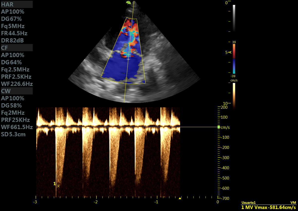
DOPPLER ECHOCARDIOGRAPHY
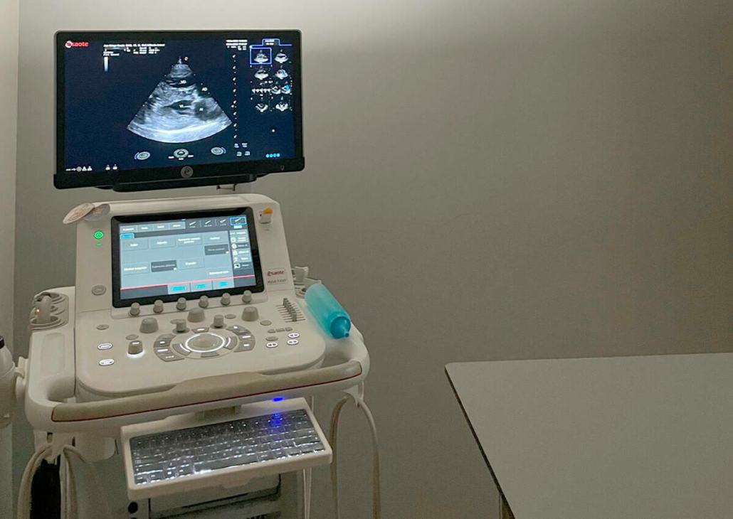
Doppler echocardiography is the imaging technique of choice for a patient with a cardiac problem.
Non-invasive technique in which information is obtained from the different intracardiac pressures, which allows an accurate diagnosis of the heart disease that is suffered and, at the same time, a prognosis of the disease in question is obtained.
With this technique we can diagnose both congenital and acquired heart conditions that our pets suffer from in our veterinary service.
1
1
Doppler echocardiography
1
1
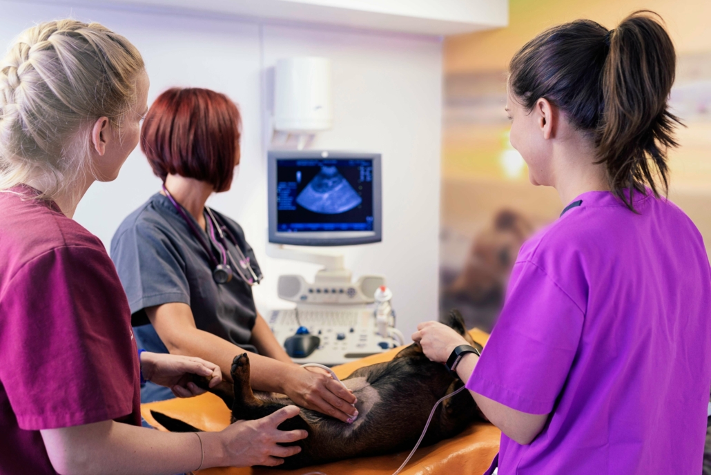
Abdominal ultrasound
ABDOMINAL ULTRASOUND
Ultrasound is the most widely used technique in veterinary medicine. It obtains an image thanks to the transmission of ultrasounds in different media.
It is a fast, non-invasive technique that allows obtaining high-resolution images and reaching a definitive diagnosis.
It allows us to evaluate the internal structure of the abdominal, thoracic, musculoskeletal and ocular organs. It is used in all specialties and can
Sampling (cytology and biopsies) of identified lesions and echo-guided loco-regional anesthesia/analgesia will also be performed.
COMPUTERIZED AXIAL TOMOGRAPHY (CT)
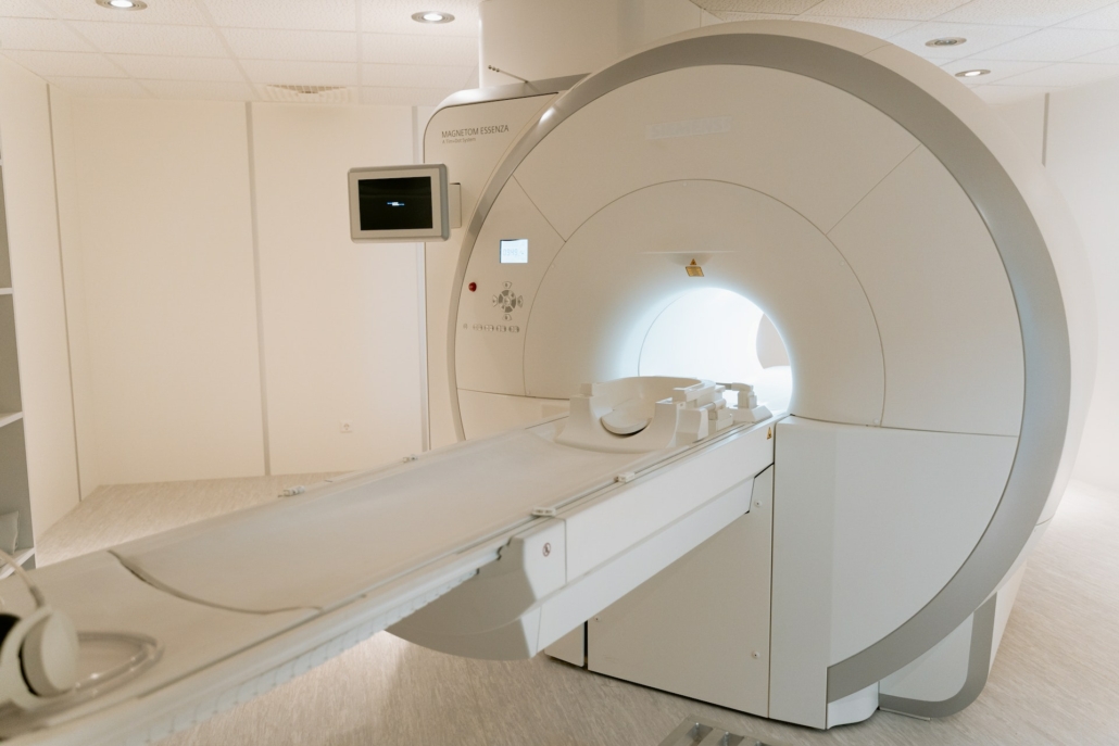
It is also based on X-rays and allows obtaining a three-dimensional image of the studied area.
It uses special X-ray equipment to create cross-sectional images of the body. To do this, a large number of images are taken from different angles, and a computer will reconstruct the images in 3D thanks to specific software.
These studies require general anesthesia since the patient must remain totally immobile while the study is performed. In certain cases, a venous contrast is injected to better visualize the desired organs.
Its main uses include cancer imaging to identify possible metastases in the thorax and abdomen, as well as delineate the margins of palpable masses.
At the cranial level and spinal cord study (mielotac) is also widely used.
1
1
CT computed tomography
1
1
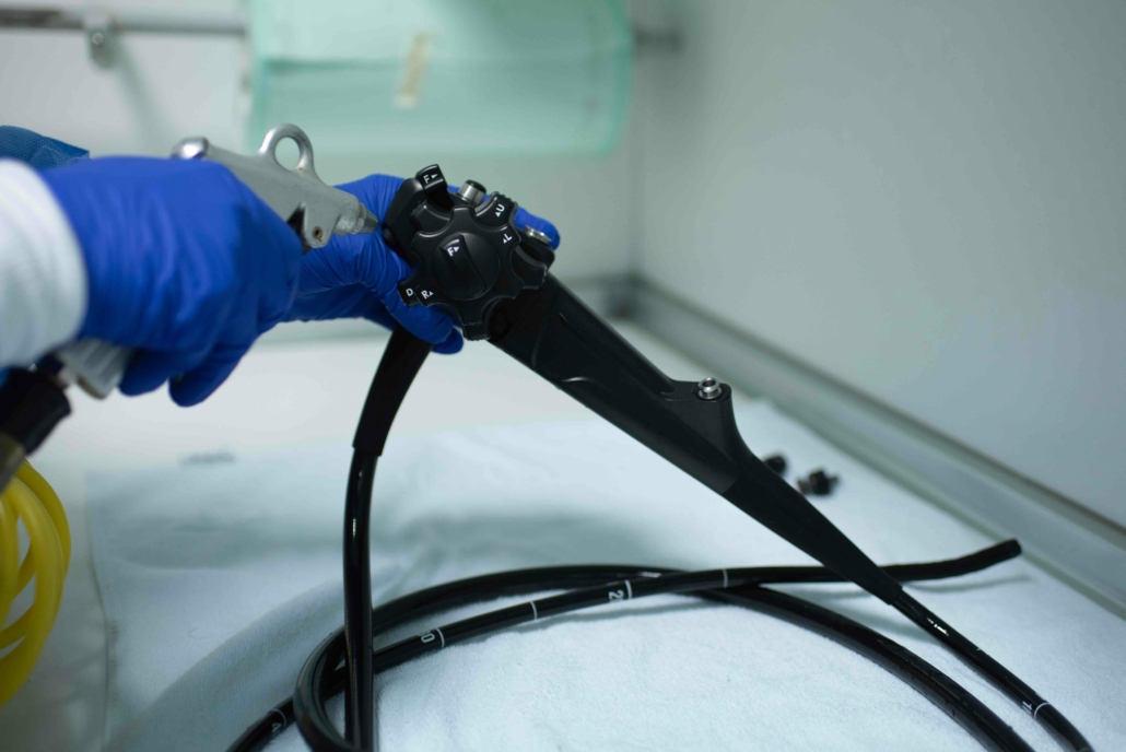
Endoscope
ENDOSCOPY
An endoscope is a long, narrow tube with a small camera at the end. General anesthesia of the patient is essential to carry out this study.
It has a large number of uses in veterinary medicine: we can examine the esophagus, stomach and duodenum. Also with this technique we can visualize the rectum and colon.
This technique allows us to take biopsies and in the event that a foreign body is observed (hook, bone…) remove it without having to perform major surgery.
It also allows us to extract plant remains (like spikes) from the nasal cavity, as well as to examine the airways of our patients and take samples (bronchoalveolar lavages) if necessary.
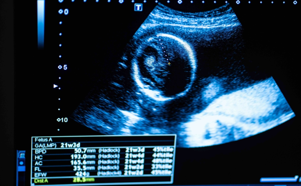
LUNG ULTRASOUND
Lung ultrasound is a non-invasive technique that allows obtaining great information about the different respiratory pathologies that small animals suffer from.
It is a technique that, by not putting stress on the patient, often allows us to approximate the diagnosis of the disease.
1
1
Veterinary lung ultrasound
ELECTROCARDIOGRAM (ECG)
Analyzes the electrical activity of the heart to study cardiac arrhythmias.
HOLTER MONITOR
It allows arrhythmias to be assessed over a longer period of time than the ECG (generally 24 hours but up to 7 days). It is a very useful tool both for diagnosing episodes of collapse, syncope and/or weakness and for assessing the control of arrhythmias in medicated patients.
BLOOD PRESSURE
Check if a patient has hypertension or hypotension. This test allows us to evaluate it in a non-invasive way.
DRAINS
Emptying of the pericardial, thoracic or abdominal cavities in cases where there are effusions.
ECHO-GUIDED PUNCTURES
Puncture and fine needle aspiration of abdominal or thoracic organs or masses for histopathological study.
AMBULANT ULTRASONOGRAPHY / DOPPLER ECHOCARDIOGRAPHY SERVICE
We went to veterinary clinics to perform abdominal ultrasound and Doppler echocardiography.
TRAINING PERIODS
Training periods are offered in ultrasound and diagnostic imaging services. For more information, write to us at our email.
+15.000
ULTRASOUND PERFORMED
+12.000
DOPPLER ULTRASOUND
+8.000
ENDOSCOPIES
+6.500
CT MADE
+20 YEARS
WITH OUR PATIENTS
54.860
DIAGNOSES MADE
100%
CUSTOMER SATISFACTION